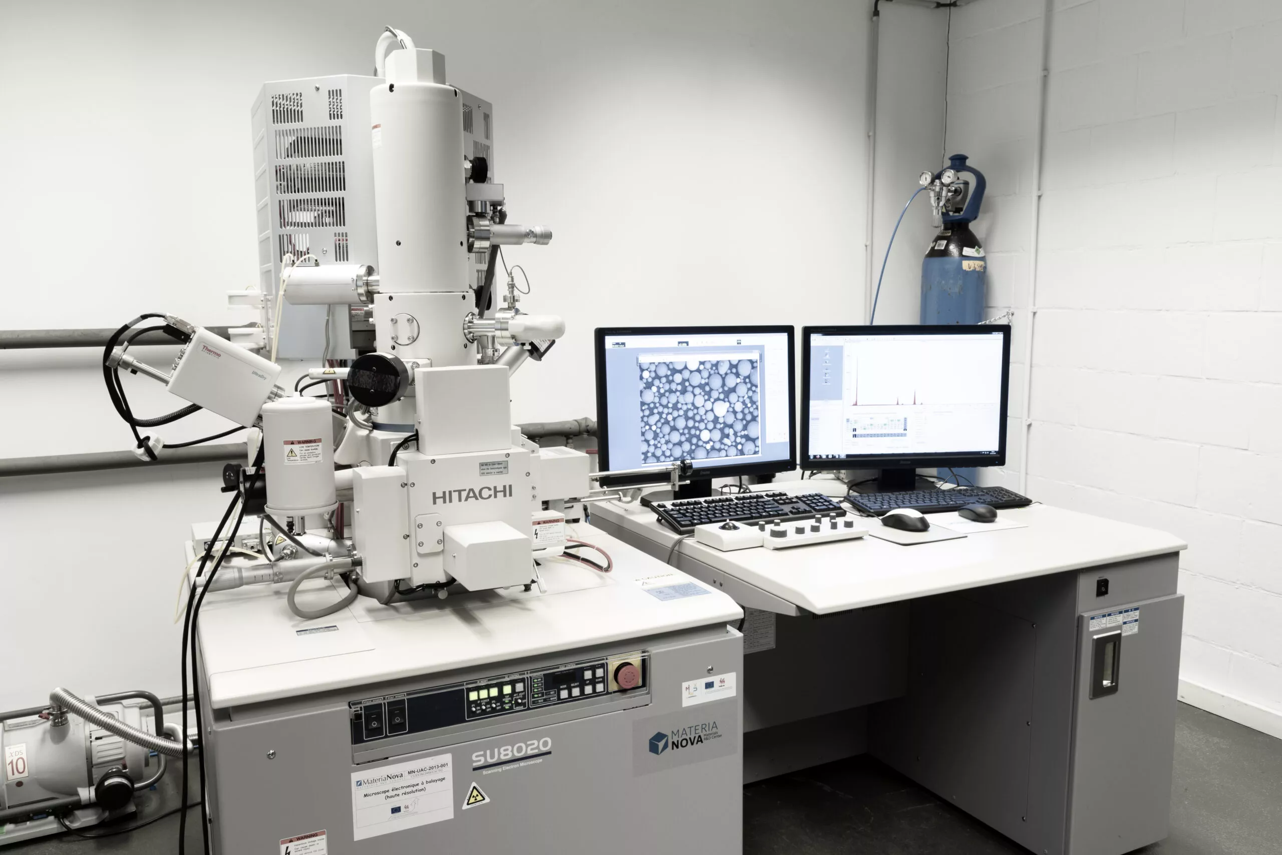
Scanning Electron Microscope (SEM)
The SEM-EDX is a microscopic analysis technique using a Field Emission Gun (FEG) which produces very high resolution images of the surface of a sample (theoretical resolution of 1 nm). This microscopic analysis tool is particularly powerful and efficient for rapid diagnosis (pollution, inclusion...) or more complex expertise. Our laboratory has the equipment to cut sections of the samples in order to carry out fine analyses on the slice (thickness measurements, multi-layer analyses, metallographic sections...).
The SEM is equipped with an EDX detector (Energy Dispersive X-ray) which allows semi-quantitative analysis of the chemical elements present. Moreover, it offers the possibility of making maps to visualize the distribution of elements.
Materia Nova has a latest generation electron microscope equipped with:
- a conventional secondary electron detector to observe the relief of the sample.
- a backscattered electron detector to detect phases of different compositions.
- an Inlens detector to obtain high-resolution images at low voltage (1.3nm resolution at 1kV), in topography or composition
- a transmitted electron detector for transmission images (STEM).
- an EDX detector to carry out elemental composition analysis.